Jaxon B. was presented to the hospital for Professional Dental Therapy on Jan 11/18. This beautiful friendly boxer had a fractured canine and periodontal disease surrounding his crowded premolars. Preanesthetic blood work was performed to ensure a safe delivery of the anesthesia. Administration of IV fluids began before he was given the anesthetic. Once anesthetized his teeth were assessed, dental radiographs were taken and the appropriate treatment was provided to his teeth. Once completed they were cleaned and polished.
The following pictures will identify the problem areas inside Jaxon’s mouth.
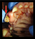
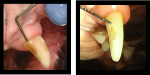
The exposed pulp chamber (small hole in the middle of the tooth indicated by the arrow) allows bacteria to enter the tooth and cause infection. The only treatment for Jaxon’s fractured upper canine tooth is an extraction. This relieved him from a painful condition and prevented a tooth abscess.
This photograph shows the sutures from Jaxon’s extraction site. The gum was cut, the bone covering the root was removed, then the tooth was extracted. These sutures will dissolve in 2 – 4 weeks.
Jaxon must have chewed on objects that were too hard and damaged the enamel of his teeth. The enamel is the white outer layer of a tooth that serves as a protective barrier. The brownish colour you are seeing is called “dentin”. This type of injury will cause affected teeth to be sensitive to hot, cold and to touch because of the exposed dentin.
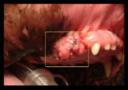
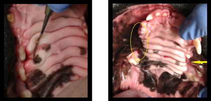
Boxers are brachycephalic dogs because of their shorter muzzle leading to crowded teeth and caused him to suffer from periodontal disease. Periodontal pockets have formed between the roots of the premolar and his bone (indicated in the photograph). The bone exposure was so serious that the same tooth on the right and the left upper jaw required extraction.
This view of Jaxon’s mouth shows, on the left side of the photograph, multiple extraction sites. (The canine tooth and a premolar were removed). The arrow on the right indicates the extraction of the affected tooth shown in the photo just above this one.
Jaxon’s remaining teeth were then cleaned and polished. A lot of debris such as food, bacteria, and some of Jaxon’s own fur were flushed out from between the folds of the hard palate. This also helped to remove a source of bad breath. At the end of the day, he was discharged from the Baxter Animal Hospital with pain medication to ensure a painless recovery.
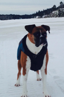
I have no doubt Jaxon feels better since the source of infection and pain no longer exists. His pet parent sent us this beautiful image of him outside getting some fresh air. She informed me that his breath is much better and he is “his usual silly self”
“Here is a picture of Jaxon enjoying a walk on the lake on a beautiful winter day. Thanks for ensuring he was at ease and taking the necessary steps to include the additional treatment during his recent dental procedure. You were all so compassionate and caring. Thank you!” – Jaxon’s Pet Parent
Written by France Filiatreault, RVT and Canadian Veterinary Dental Technician